This article was written using specialized medical literature. All material used has been analyzed and presented in easy-to-understand language with minimal use of medical terms. The purpose of this article was an accessible explanation of the meaning of a general blood test and interpretation of its results.
If you have identified a deviation from the norm in a general blood test and want to learn more about the possible causes, then click on the selected blood value in the table - this will allow you to go to the selected section.
The article provides detailed information about the norms of cellular elements for each age. Deciphering a blood test in children requires special attention. Normal blood levels in children depend on age, so accurate information about the child's age is necessary to interpret the results of a blood test. You can find out about age norms from the tables below - separate for each blood test indicator.
We all have had a general blood test at least once in our lives. And every person was faced with a misunderstanding about what was written on the form, what did all these numbers mean? How to understand why this or that indicator is increased or decreased? What could be the risk of an increase or decrease, for example, in lymphocytes? Let's look at everything in order.
General blood test norms
| Analysis indicator | Norm |
| Hemoglobin | Men: 130-170 g/l |
| Women: 120-150 g/l | |
| Red blood cell count | Men: 4.0-5.0 10 12 /l |
| Women: 3.5-4.7 10 12 /l | |
| White blood cell count | Within 4.0-9.0x10 9 /l |
| Hematocrit (the ratio of the volume of plasma and cellular elements of blood) | Men: 42-50% |
| Women: 38-47% | |
| Average red blood cell volume | Within 86-98 microns 3 |
| Leukocyte formula | Neutrophils:
Monocytes: 3-11% Eosinophils: 0.5-5% Basophils: 0-1% |
| Platelet count | Within 180-320 10 9 /l |
| Erythrocyte sedimentation rate (ESR) | Men: 3 - 10 mm/h |
| Women: 5 - 15 mm/h |
Hemoglobin
| Hemoglobin (Hb) is a protein containing an iron atom that is capable of attaching and transporting oxygen. Hemoglobin is found in red blood cells. The amount of hemoglobin is measured in grams/liter (g/l). Determining the amount of hemoglobin is very important, since when its level decreases, the tissues and organs of the entire body experience a lack of oxygen. | |||||||||||||||||||||||||||||||||||||||||||||||||||||||||||
| Reasons for increased hemoglobin
|
||||||||||||||||||||||||||||||||||||||||||||||||||||||||||
Low hemoglobin - reasons
|
|||||||||||||||||||||||||||||||||||||||||||||||||||||||||||
Red blood cell count
| Red blood cells- These are small red blood cells. These are the most numerous blood cells. Their main function is the transfer of oxygen and its delivery to organs and tissues. Red blood cells are presented in the form of biconcave discs. Inside the red blood cell there is a large amount of hemoglobin - the main volume of the red disk is occupied by it. | ||||||||||||||||||||||||||||||||
| Causes of decreased red blood cell levelsA decrease in the number of red blood cells is called anemia. There are many reasons for the development of this condition, and they are not always associated with the hematopoietic system.
|
|||||||||||||||||||||||||||||||
Reasons for the increase in the number of red blood cells
|
||||||||||||||||||||||||||||||||
| What to do if red blood cells are elevated? | ||||||||||||||||||||||||||||||||
Total white blood cell count
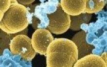
| Leukocytes- these are living cells of our body circulating with the bloodstream. These cells carry out immune control. In the event of an infection or damage to the body by toxic or other foreign bodies or substances, these cells fight the damaging factors. The formation of leukocytes occurs in the red bone marrow and lymph nodes. Leukocytes are divided into several types: neutrophils, basophils, eosinophils, monocytes, lymphocytes. Different types of leukocytes differ in appearance and functions performed during the immune response. | |
Causes of increased leukocytesPhysiological increase in leukocyte levels
|
|
Causes of decreased leukocytes
|
|
Hematocrit
| Hematocrit– this is the percentage ratio of the volume of the blood being tested to the volume occupied by red blood cells in it. This indicator is calculated as a percentage. | ||||||||||||||||||||||||||||||||||||||||||||||||||||||||||||||
| Reasons for increased hematocrit
|
|||||||||||||||||||||||||||||||||||||||||||||||||||||||||||||
Reasons for decreased hematocrit
|
||||||||||||||||||||||||||||||||||||||||||||||||||||||||||||||
MCH, MCHC, MCV, color index (CPU)- norm
Color Index (CPU)- This is a classic method for determining the hemoglobin concentration in red blood cells. Currently, it is gradually being replaced by the MCH index in blood tests. These indices reflect the same thing, only expressed in different units.
Leukocyte formula
The leukocyte formula is an indicator of the percentage of different types of leukocytes in the blood and the total number of leukocytes in the blood (this indicator is discussed in the previous section of the article). The percentage of different types of leukocytes in infectious, blood diseases, and oncological processes will change. Thanks to this laboratory symptom, the doctor may suspect the cause of health problems.Types of leukocytes, normal
| Neutrophils | Segmented forms 47-72% |
| Band forms 1-6% | |
| Eosinophils | 0,5-5% |
| Basophils | 0-1% |
| Monocytes | 3-11% |
| Lymphocytes | 19-37% |
In order to find out the age norm, click on the name of the leukocyte from the table.
Neutrophils
| Neutrophils There can be two types - mature forms, which are also called segmented, and immature - rod-shaped. Normally, the number of band neutrophils is minimal (1-3% of the total number). With the “mobilization” of the immune system, there is a sharp increase (by several times) in the number of immature forms of neutrophils (band neutrophils). | ||||||||||||||||||||||||||||||||||
| An increase in the level of neutrophils in the blood is a condition called neutrophilia. Reasons for increased neutrophil levels
|
|||||||||||||||||||||||||||||||||
| Decreased neutrophil levels - a condition called neutropenia Reasons for decreased neutrophil levels
|
||||||||||||||||||||||||||||||||||
What is a shift in the leukocyte formula to the left and to the right?Shift of the leukocyte formula to the left means that young, “immature” neutrophils appear in the blood, which are normally present only in the bone marrow, but not in the blood. A similar phenomenon is observed in mild and severe infectious and inflammatory processes (for example, sore throat, malaria, appendicitis), as well as in acute blood loss, diphtheria, pneumonia, scarlet fever, typhus, sepsis, intoxication.Shift of the leukocyte formula to the right means that the number of “old” neutrophils (segmented) in the blood increases, and the number of nuclear segments becomes more than five. This picture occurs in healthy people living in areas contaminated with radiation waste. It is also possible in the presence of B 12 deficiency anemia, with a lack of folic acid, in people with chronic lung disease, or with obstructive bronchitis. |
||||||||||||||||||||||||||||||||||
Eosinophils
| Eosinophils– this is one of the types of leukocytes that are involved in cleansing the body of toxic substances, parasites, and participates in the fight against cancer cells. This type of leukocyte is involved in the formation of humoral immunity (immunity associated with antibodies) | |
Causes of increased blood eosinophils
|
|
Reasons for the decrease in eosinophils
|
|
Monocytes
| Monocytes- few, but the largest immune cells in the body. These white blood cells are involved in recognizing foreign substances and training other white blood cells to recognize them. They can migrate from the blood into body tissues. Outside the bloodstream, monocytes change their shape and transform into macrophages. Macrophages can actively migrate to the site of inflammation in order to take part in cleansing the inflamed tissue of dead cells, leukocytes, and bacteria. Thanks to this work of macrophages, all conditions are created for the restoration of damaged tissues. | |
Causes of increased monocytes (monocytosis)
|
|
Causes of decreased monocytes (monocytopenia)
|
|
Basophils
Causes of increased blood basophils
- decreased thyroid hormone levels hypothyroidism
- chicken pox
- food and drug allergies
- condition after removal of the spleen
- treatment with hormonal drugs (estrogens, drugs that reduce the activity of the thyroid gland)
Lymphocytes
| Lymphocytes– the second largest fraction of leukocytes. Lymphocytes play a key role in humoral (through antibodies) and cellular (implemented through direct contact of the destroyed cell and lymphocyte) immunity. Different types of lymphocytes circulate in the blood - helpers, suppressors and killers. Each type of leukocyte is involved in the formation of the immune response at a certain stage. | |
Causes of increased lymphocytes (lymphocytosis)
|
|
Causes of low lymphocytes (lymphopenia)
|
|
Platelets
Causes of increased platelets
(thrombocytosis, platelet count more than 320x10 9 cells/l)- splenectomy
- inflammatory processes (exacerbation of rheumatism,
A general blood test is perhaps the most common laboratory diagnostic method. In modern civilized society, there is practically not a single person who does not have to repeatedly donate blood for a general analysis.
After all, this study is carried out not only on sick people, but also on completely healthy people during routine medical examinations at work, in educational institutions, and in the army.
And for various diseases, a general blood test is mandatory and is included in the standard of any clinical research.
Hematocrit– this is the percentage ratio of formed elements, dry residue to the total volume of blood. This dry residue is mainly represented by red blood cells - the influence of other formed elements on the hematocrit is not significant due to their relatively low content.
Normally, in men the hematocrit is in the range of 39–49%, in women – 35–45%.
A decrease in hematocrit is most often due to blood loss, and an increase indicates blood thickening. The color indicator is the degree of saturation of the red blood cell with hemoglobin. Normally it is from 0.85 to 1.15. This indicator decreases in hypochromic iron deficiency anemia.
Leukocytes
Leukocytes are white blood cells. The main function of leukocytes is to protect the body from infection, pathological external influences, and neutralize various toxins.
In 1l. blood contains from 4 to 9 X 10 9 leukocytes.
An increase in the number of leukocytes (leukocytosis) is observed in many pathological conditions - infections, poisoning, injuries, diseases of internal organs, after blood loss and surgical interventions. Leukocytosis is also observed during pregnancy, after heavy fatty foods and physical activity. A decrease in the number of leukocytes (leukopenia) is observed in weakened and exhausted patients, after long-term use of certain medications. Leukopenia indicates low body resistance and the danger of infectious diseases.
Leukocytes are not homogeneous in composition. The percentage of their varieties is displayed in the so-called. leukocyte formula.
- Eosinophils 0-5
- Basophils 0-1
- Neutrophils
- Band 1-5
- Segmented 47-72
- Lymphocytes 21-38
- Monocytes 4-10
All leukocytes are divided into 2 types - granulocytes and agranulocytes.
Granulocytes have a specific granularity in their cytoplasm. This granularity can be stained with acidic (eosinophils), basic (basophils) and neutral (neutrophils) dyes.
In agranulocytes (lymphocytes, monocytes) such granularity is absent.
An increase in the level of eosinophils is observed in helminthic infestations, tuberculosis and various allergic conditions, including bronchial asthma. The absence of eosinophils (aneosinophilia) is detected in infectious diseases, anemia, severe injuries, and after surgery. The number of basophils has no significant clinical significance.
Neutrophils– the most numerous (in adults) type of leukocytes. Their function is to neutralize microbial cells and foreign particles by phagocytosis. Neutrophils themselves can be mature (segmented) and maturing (band). An increase in the number of neutrophils is observed during infections, mainly bacterial, injuries, myocardial infarction, and malignant tumors. In severe diseases, mainly band neutrophils increase - the so-called. rod shift to the left. In particularly severe conditions, purulent processes and sepsis, young forms can be detected in the blood - promyelocytes and myelocytes, which should not normally be present. Also, during severe processes, toxic granularity is detected in neutrophils.
An increase in the level of lymphocytes is observed in viral infections - influenza, viral hepatitis, rubella, as well as in tumors of the hematopoietic organs. The function of monocytes is phagocytosis. They increase with tuberculosis, syphilis, rheumatism, and diseases of the hematopoietic organs. The reasons for the decrease in the level of agranulocytes (lymphocytes and monocytes) are severe illnesses leading to exhaustion of the patient, long-term use of certain medications.
Platelets
These are blood platelets that help blood clot and stop bleeding (hemostasis).
Normally 1 liter. blood contains from 200 to 300x10 9.
A decrease in this indicator (thrombocytopenia) is observed with viral and bacterial infections, after blood loss and massive injuries, with some connective tissue diseases, and with bone marrow tumors.
Thrombocytopenia is a dangerous sign indicating the risk of massive bleeding.
An increase in platelets (thrombocytosis) develops after removal of the spleen, surgical interventions, and malignant tumors. Thrombocytosis may also be secondary after hemodilution. The main danger of thrombocytosis is thrombosis, intravascular coagulation of blood, leading to severe damage to organs and tissues. It should be noted that the level of platelets in a general blood test does not provide a comprehensive picture of blood coagulation. This requires another blood test - a coagulogram.
Conclusion
In conclusion, it should be noted that the data of a general blood test are mostly nonspecific. And based on this study alone, it is unlikely that a diagnosis can be made. Existing deviations serve as a reason for a more in-depth diagnosis. In addition, the norms of general analysis vary too much between both sexes and different age categories. This can be seen in the example of children, whose normal blood picture may differ significantly from that of adults. And the standards themselves are revised from time to time by clinicians and laboratory assistants. Therefore, in different sources you can find meanings that are slightly different from each other.
We try to provide the most relevant and useful information for you and your health. The materials posted on this page are informational in nature and intended for educational purposes. Site visitors should not use them as medical advice. Determining the diagnosis and choosing a treatment method remains the exclusive prerogative of your attending physician! We are not responsible for possible negative consequences arising from the use of information posted on the website
A general blood test is perhaps the most common laboratory diagnostic method. In modern civilized society, there is practically not a single person who does not have to repeatedly donate blood for a general analysis.
After all, this study is carried out not only on sick people, but also on completely healthy people during routine medical examinations at work, in educational institutions, and in the army.
This blood test includes determining the concentration of hemoglobin, the number of leukocytes and counting the leukocyte formula, determining the number of red blood cells, platelets, erythrocyte sedimentation rate (ESR) and other indicators.
Thanks to the correct interpretation of the results of a general blood test, it is possible to establish the cause of certain symptoms in adults, determine the type of disease of the blood and internal organs, and select the correct treatment regimen.
What is it?
A general (detailed) blood test includes:
- Hemoglobin and hematocrit levels.
- Erythrocyte sedimentation rate (ESR), formerly called reaction rate (ERR).
- Color index calculated according to the formula, if the study was carried out manually, without the participation of laboratory equipment;
- Determination of the content of cellular elements of blood: erythrocytes - red blood cells containing the pigment hemoglobin, which determines the color of blood, and leukocytes, which do not contain this pigment, therefore are called white blood cells (neutrophils, eosinophils, basophils, lymphocytes, monocytes).
As you can see, a general blood test shows the reaction of this valuable biological fluid to any processes occurring in the body. Regarding correct analysis, then there are no complex, strict regulations regarding this testing, but there are certain restrictions:
- The analysis is carried out in the morning. The patient is prohibited from consuming food or water 4 hours before taking a blood sample.
- The main medical supplies used for drawing blood are a scarifier, cotton wool, and alcohol.
- For this examination, capillary blood is used, which is taken from a finger. Less often, according to the doctor's instructions, blood from a vein can be used.
After receiving the results, a detailed breakdown of the blood test is performed. There are also special hematology analyzers that can automatically determine up to 24 blood parameters. These devices are capable of producing a printout with a transcript of the blood test almost immediately after blood collection.
Complete blood count: normal indicators in the table
The table shows the normal number of blood elements. These values may differ in different laboratories, so to find out whether the blood test results accurately correspond to the norm, it is necessary to find out the reference values of the laboratory in which the blood test was carried out.
Table of normal indicators of a general blood test in adults:
| Analysis: | Adult women: | Adult men: |
| Hemoglobin | 120-140 g/l | 130-160 g/l |
| Hematocrit | 34,3-46,6% | 34,3-46,6% |
| Platelets | 180-360×109 | 180-360×109 |
| Red blood cells | 3.7-4.7×10¹² | 4-5.1×10¹² |
| Leukocytes | 4-9×10⁹ | 4-9×10⁹ |
| ESR | 2-15 mm/h | 1-10 mm/h |
| Color index | 0,85-1,15 | 0,85-1,15 |
| Reticulocytes | 0,2-1,2% | 0,2-1,2% |
| Thrombocrit | 0,1-0,5% | 0,1-0,5% |
| Eosinophils | 0-5% | 0-5% |
| Basophils | 0-1% | 0-1% |
| Lymphocytes | 18-40% | 18-40% |
| Monocytes | 2-9% | 2-9% |
| Average red blood cell volume | 78-94 fl | 78-94 fl |
| Average hemoglobin content in erythrocytes | 26-32 pg | 26-32 pg |
| Band granulocytes (neutrophils) | 1-6% | 1-6% |
| Segmented granulocytes (neutrophils) | 47-72% | 47-72% |
Each of the given indicators is important when deciphering a blood test, however, a reliable result of the study consists not only of comparing the data obtained with the norms - all quantitative characteristics are considered together, in addition, the relationship between various indicators of blood properties is taken into account.
Red blood cells
Formed elements of blood. They contain hemoglobin, which is found in each of the red blood cells in equal quantities. Red blood cells transport oxygen and carbon dioxide in the body.
Promotion:
- Vaquez disease (erythremia) is a chronic leukemia.
- As a result of hypohydration with sweating, vomiting, burns.
- As a consequence of hypoxia in the body in chronic diseases of the lungs, heart, narrowing of the renal arteries and polycystic kidney disease. An increase in erythropoietin synthesis in response to hypoxia leads to an increase in the formation of red blood cells in the bone marrow.
Decline:
- Anemia.
- Leukemia, myeloma - blood tumors.
The level of red blood cells in the blood also becomes lower in diseases that are characterized by increased breakdown of red blood cells:
- hemolytic anemia;
- iron deficiency in the body;
- lack of vitamin B12;
- bleeding.
The average lifespan of an erythrocyte is 120 days. These cells are formed in the bone marrow and destroyed in the liver.
Platelets
Formed elements of blood involved in ensuring hemostasis. Platelets are formed in the bone marrow from megakaryocytes.
An increase in the number of platelets (thrombocytosis) is observed when:
- bleeding;
- splenectomy;
- reactive thrombocytosis;
- treatment with corticosteroids;
- physical stress;
- iron deficiency;
- malignant neoplasms;
- acute hemolysis;
- myeloproliferative disorders (erythremia, myelofibrosis);
- chronic inflammatory diseases (rheumatoid arthritis, tuberculosis, cirrhosis of the liver).
A decrease in platelet count (thrombocytopenia) is observed with:
- decreased platelet production;
- DIC syndrome;
- increased destruction of platelets;
- hemolytic-uremic syndrome;
- splenomegaly;
- autoimmune diseases.
The main function of this blood component is to participate in blood clotting. Platelets contain the bulk of clotting factors, which are released into the blood when necessary (damage to the vessel wall). Thanks to this property, the damaged vessel is clogged by the forming thrombus and bleeding stops.

Leukocytes
White blood cells. Formed in red bone marrow. The function of leukocytes is to protect the body from foreign substances and microbes. In other words, it is immunity.
Increase in leukocytes:
- infections, inflammation;
- allergy;
- leukemia;
- condition after acute bleeding, hemolysis.
Decrease in leukocytes:
- bone marrow pathology;
- infections (flu, rubella, measles, etc.);
- genetic abnormalities of immunity;
- increased spleen function.
There are different types of leukocytes, so a change in the number of individual types, and not all leukocytes in general, is of diagnostic importance.
Basophils
When released into the tissues, they turn into mast cells, which are responsible for the release of histamine - a hypersensitivity reaction to food, medications, etc.
- Increased: hypersensitivity reactions, chicken pox, hypothyroidism, chronic sinusitis.
- Decreased: hyperthyroidism, pregnancy, ovulation, stress, acute infections.
Basophils take part in the formation of delayed-type immunological inflammatory reactions. They contain large amounts of substances that cause tissue inflammation.
Eosinophils
Cells that are responsible for allergies. Normally, they should be from 0 to 5%. If the indicator increases, it indicates the presence of allergic inflammation (allergic rhinitis). It is important that the number of eosinophils can be increased in the presence of helminthic infestations! This happens especially often in children. This fact should be taken into account by pediatricians to make the correct diagnosis.
Neutrophils
They are divided into several groups - young, rod and segmented. Neutrophils provide antibacterial immunity, and their varieties are the same cells of different ages. Thanks to this, it is possible to determine the severity and severity of the inflammatory process or damage to the hematopoietic system.
An increase in the number of neutrophils is observed during infections, mainly bacterial, injuries, myocardial infarction, and malignant tumors. In severe diseases, mainly band neutrophils increase - the so-called. rod shift to the left. In particularly severe conditions, purulent processes and sepsis, young forms can be detected in the blood - promyelocytes and myelocytes, which should not normally be present. Also, during severe processes, toxic granularity is detected in neutrophils.

MON - monocytes
This element is considered a variation of leukocytes in the macrophage form, i.e. their active phase, absorbing dead cells and bacteria. The norm for a healthy person is from 0.1 to 0.7 * 10^9 e/l.
A decrease in MON levels is due to severe operations and the use of corticosteroids; an increase indicates the development of rheumatoid arthritis, syphilis, tuberculosis, mononucleosis and other diseases of an infectious nature.
GRAN - granulocytes
Granulated leukocytes are activators of the immune system in the fight against inflammation, infections and allergic reactions. The norm for humans is from 1.2 to 6.8 * 10^9 e/l.
GRAN levels increase in inflammation and decrease in lupus erythematosus and aplastic anemia.
Color index
Reflects the relative content of hemoglobin in red blood cells. Used for the differential diagnosis of anemia: normochromic (normal amount of hemoglobin in the red blood cell), hyperchromic (increased), hypochromic (decreased).
- A decrease in CP occurs with: iron deficiency anemia; anemia caused by lead intoxication, in diseases with impaired hemoglobin synthesis.
- An increase in CP occurs with: insufficiency of vitamin B12 in the body; folic acid deficiency; cancer; polyposis of the stomach.
Color index (CI): 0.85-1.1.
Hemoglobin
An increase in hemoglobin concentration occurs with erythremia (a decrease in the number of red blood cells), erythrocytosis (an increase in the number of red blood cells), as well as with blood thickening - a consequence of a large loss of fluid by the body. In addition, the hemoglobin level is increased with cardiovascular decompensation.
If the hemoglobin value is more or less than the normal limit, this indicates the presence of pathological conditions. Thus, a decrease in the concentration of hemoglobin in the blood is observed with anemia of various etiologies and with blood loss. This condition is also called anemia.
Hematocrit
Hematocrit is the percentage ratio of the volume of the blood being tested to the volume occupied by red blood cells in it. This indicator is calculated as a percentage.
A decrease in hematocrit occurs when:
- anemia;
- fasting;
- pregnancy;
- water retention in the body (chronic renal failure);
- excess protein content in plasma (myeloma);
- drinking plenty of fluids or administering large amounts of intravenous solutions.
An increase in hematocrit above normal indicates:
- leukemia;
- polycythemia vera;
- burn disease;
- diabetes mellitus;
- kidney diseases (hydronephrosis, polycystic disease, neoplasms);
- fluid loss (excessive sweating, vomiting);
- peritonitis.
Normal hematocrit values: Men – 40-48%, women – 36-42%.

ESR
The erythrocyte sedimentation rate shows how quickly the blood separates into two layers - the upper (plasma) and lower (formed elements). This indicator depends on the number of red blood cells, globulins and fibrinogen. That is, the more red cells a person has, the slower they settle. An increase in the amount of globulins and fibrinogen, on the contrary, accelerates erythrocyte sedimentation.
Reasons for high ESR in a general blood test:
- Acute and chronic inflammatory processes of infectious origin (pneumonia, rheumatism, syphilis, tuberculosis, sepsis).
- Heart damage (myocardial infarction – damage to the heart muscle, inflammation, synthesis of “acute phase” proteins, including fibrinogen.)
- Diseases of the liver (hepatitis), pancreas (destructive pancreatitis), intestines (Crohn's disease, ulcerative colitis), kidneys (nephrotic syndrome).
- Hematological diseases (anemia, lymphogranulomatosis, myeloma).
- Endocrine pathology (diabetes mellitus, thyrotoxicosis).
- Injury to organs and tissues (surgeries, wounds and bone fractures) - any damage increases the ability of red blood cells to aggregate.
- Conditions accompanied by severe intoxication.
- Lead or arsenic poisoning.
- Malignant neoplasms.
An ESR below normal is typical for the following body conditions:
- Obstructive jaundice and, as a consequence, the release of large amounts of bile acids;
- High levels of bilirubin (hyperbilirubinemia);
- Erythremia and reactive erythrocytosis;
- Sickle cell anemia;
- Chronic circulatory failure;
- Decreased fibrinogen levels (hypofibrinogenemia).
ESR, as a nonspecific indicator of the disease process, is often used to monitor its progress.
General blood test, perhaps the most common test that doctors prescribe in order to correctly diagnose and conduct a study of the patient’s health status. But what comes in the answer does not tell the patient anything; to understand what all these numbers mean, we provide you interpretation of blood test values.
A general blood test is divided into:
- Biochemical blood test;
- Immunological blood test;
- Hormonal blood test;
- Serological blood tests.
Blood test interpretation:
|
Designations reductions |
Normal values - complete blood count | ||||||||
| children aged | adults | ||||||||
| 1 day | 1 month | 6 months | 12 months | 1-6 years | 7-12 years | 13-15 years old | man | woman | |
| Hemoglobin Hb, g/l |
180-240 | 115-175 | 110-140 | 110-135 | 110-140 | 110-145 | 115-150 | 130-160 | 120-140 |
| Red blood cells R.B.C. |
4,3-7,6 | 3,8-5,6 | 3,5-4,8 | 3,6-4,9 | 3,5-4,5 | 3,5-4,7 | 3,6-5,1 | 4-5,1 | 3,7-4,7 |
| Color index MCHC, % |
0,85-1,15 | 0,85-1,15 | 0,85-1,15 | 0,85-1,15 | 0,85-1,15 | 0,85-1,15 | 0,85-1,15 | 0,85-1,15 | 0,85-1,15 |
| Reticulocytes RTC |
3-51 | 3-15 | 3-15 | 3-15 | 3-12 | 3-12 | 2-11 | 0,2-1,2 | 0,2-1,2 |
| Platelets PLT |
180-490 | 180-400 | 180-400 | 180-400 | 160-390 | 160-380 | 160-360 | 180-320 | 180-320 |
| ESR ESR |
2-4 | 4-8 | 4-10 | 4-12 | 4-12 | 4-12 | 4-15 | 1-10 | 2-15 |
| Leukocytes WBC, % |
8,5-24,5 | 6,5-13,8 | 5,5-12,5 | 6-12 | 5-12 | 4,5-10 | 4,3-9,5 | 4-9 | 4-9 |
| Bands, % | 1-17 | 0,5-4 | 0,5-4 | 0,5-4 | 0,5-5 | 0,5-5 | 0,5-6 | 1-6 | 1-6 |
| Segmented, % | 45-80 | 15-45 | 15-45 | 15-45 | 25-60 | 35-65 | 40-65 | 47-72 | 47-72 |
| Eosinophils EOS, % |
0,5-6 | 0,5-7 | 0,5-7 | 0,5-7 | 0,5-7 | 0,5-7 | 0,5-6 | 0-5 | 0-5 |
| Basophils BAS, % |
0-1 | 0-1 | 0-1 | 0-1 | 0-1 | 0-1 | 0-1 | 0-1 | 0-1 |
| Lymphocytes LYM, % |
12-36 | 40-76 | 42-74 | 38-72 | 26-60 | 24-54 | 25-50 | 18-40 | 18-40 |
| Monocytes MON, % |
2-12 | 2-12 | 2-12 | 2-12 | 2-10 | 2-10 | 2-10 | 2-9 | 2-9 |
Now, more about the main indicators of a general blood test.
Hemoglobin
Hemoglobin is the blood pigment of red blood cells. Its function is to transfer oxygen from the lungs to tissues and organs, and carbon dioxide back to the lungs.
Hemoglobin increase:
- staying at high altitudes
- polycythemia (increased red blood cell count)
- dehydration and blood thickening
Decreased hemoglobin:
- anemia
Color index
The color index shows the relative content of hemoglobin in red blood cells. This indicator is important in diagnosing anemia.
Increase in color index:
- spherocytosis
Reduced color index:
- iron deficiency anemia
Red blood cells
Red blood cells are red blood cells that are produced in the red bone marrow. Red blood cells contain hemoglobin and carry oxygen.
Increase in red blood cells:
- dehydration
- polycythemia
Decreased red blood cells:
- anemia
Leukocytes
White blood cells. Formed in red bone marrow. The function of leukocytes is to protect the body from foreign substances and microbes. In other words, it is immunity.
There are different types of leukocytes, so a change in the number of individual types, and not all leukocytes in general, is of diagnostic importance.
Increase in leukocytes:
- infections, inflammation
- allergy
- leukemia
- condition after acute bleeding, hemolysis
Decrease in leukocytes:
- bone marrow pathology
- infections (flu, rubella, measles, etc.)
- genetic abnormalities of immunity
- increased spleen function
Leukocyte formula
Percentage of different types of leukocytes. Neutrophils: cells responsible for inflammation, fighting infection (except viral ones), nonspecific defense (immunity), removing one’s own dead cells. Mature neutrophils have a segmented nucleus, while young ones have a rod-shaped nucleus.
Increased leukocyte count:
- intoxication
- infections
- inflammatory process
- malignant tumors
- psycho-emotional arousal
Decrease in leukocyte formula:
- aplastic anemia, bone marrow pathology
- genetic immune disorders
- some infections (viral, chronic)
Eosinophils
Decrease in eosinophils:
- purulent infections
- surgery
Basophils
When basophils enter the tissues, they turn into mast cells, which are responsible for the release of histamine - a hypersensitivity reaction to food, drugs, etc.
Increased basophils:
- chicken pox
- hypersensitivity reactions
- chronic sinusitis
- hypothyroidism
Decrease in basophils:
- pregnancy
- ovulation
- acute infections
- hyperthyroidism
- stress
Lymphocytes
Lymphocytes are the main cells of the human body's immune system. They fight viral infections, destroy foreign cells and altered own cells, and release antibodies (immunoglobulins) into the blood - substances that block antigen molecules and remove them from the body.
Increased lymphocytes:
- lymphocytic leukemia
- viral infections
Decreased lymphocytes:
- loss of lymph
- aplastic anemia
- acute infections (non-viral) and diseases
- immunodeficiency states
- systemic lupus erythematosus
Monocytes
Monocytes are the largest white blood cells. They finally destroy foreign cells and proteins, foci of inflammation, and destroyed tissues. Monocytes are the most important cells of the immune system; it is monocytes that first encounter antigen and present it to lymphocytes for the development of a full immune response.
Increased monocytes:
- leukemia
- tuberculosis, sarcoidosis, syphilis
- infections (viral, fungal, protozoal)
- systemic connective tissue diseases (arthritis, periarteritis nodosa, systemic lupus erythematosus)
Decrease in monocytes:
- hairy cell leukemia
- aplastic anemia
ESR
ESR is the erythrocyte sedimentation rate when blood settles. The ESR level depends directly on the number of red blood cells, their “weight” and shape, as well as on the properties of the blood plasma - the amount of proteins, as well as viscosity.
Increasing ESR:
- inflammatory process
- infections
- anemia
- malignant tumors
- pregnancy
Reticulocytes
Reticulocytes are young forms of red blood cells. Normally they should be located in the bone marrow. Their excess blood output indicates an increased rate of red blood cell formation.
Increased reticulocytes:
- increased formation of red blood cells in anemia (blood loss, iron deficiency, hemolytic)
Decrease in reticulocytes:
- kidney diseases
- disorders of red blood cell maturation (B12 folate deficiency anemia)
- aplastic anemia
Platelets
Platelets are platelets of blood that are formed from giant cells in the bone marrow. Platelets are responsible for blood clotting.
Increase in platelets:
- inflammatory process
- myeloid leukemia
- polycythemia
- condition after surgery
Decreased platelets:
- aplastic anemia
- systemic lupus erythematosus
- thrombocytopenic purpura
- hemolytic disease, isoimmunization by blood groups, Rh factor
- hemolytic anemia
However, it is worth remembering that only a doctor can make a correct diagnosis and interpret the tests. All of the above is for guidance only, but not for independent diagnosis.
The survey method under consideration is one of the most popular. Thanks to a general blood test, it is possible to establish the cause of certain symptoms, determine the type of disease of the blood and internal organs, and select adequate treatment. To correctly interpret this analysis, you need to know the normal blood parameters.
How to take a general blood test and what is needed for this?
There are no complex, strict regulations regarding this testing, but there are some rules:
- For this examination, capillary blood is used, which is taken from a finger. Less often, according to the doctor's instructions, blood from a vein can be used.
- The analysis is carried out in the morning. The patient is prohibited from consuming food or water 4 hours before taking a blood sample.
- The main medical supplies used for drawing blood are a scarifier, cotton wool, and alcohol.
The algorithm for collecting capillary blood is as follows:
- The finger from which blood is planned to be drawn is treated with alcohol. For better blood sampling, it is useful to pre-rub your finger to ensure better blood flow to it.
- A scarifier is used to pierce the skin of the finger.
- Blood is collected using a small pipette. The sample is placed in a sterile tube.
Deciphering the general blood test of a child and an adult, the norms in the tables and the reasons for deviations from the norms.
Everyone in their life has gone through such a painless procedure as donating blood from a finger. But for most, the result obtained remains just a set of numbers written on paper. Explanations of this analysis will enable each patient to navigate the deviations that were detected in the blood and the reasons that caused them.
General blood test - hemoglobin content in the blood.
This blood component is a protein, through which oxygen is supplied to all internal organs/systems. The amount of this component is calculated in grams, which is in 1 liter of blood.
- Norms of hemoglobin content in the blood of children and adults.
This indicator will depend on the patient’s age and gender:

- Causes of increased and decreased hemoglobin levels in children and adults.
An increased level of hemoglobin is observed with:
- Diagnosis.
- Kidney diseases.
- The patient has pathologies associated with hematopoiesis.
Low hemoglobin levels may result from:
- Vitamin/iron deficiency.
- Significant blood loss.
- Blood cancer.
- Anemia.
- A strict diet that led to exhaustion.
Red blood cells in a general blood test.
The components in question contain hemoglobin. The main purpose of red blood cells is to carry oxygen to internal organs. Often in the table, instead of the unit of measurement of red blood cells, you can see the abbreviation RBC.

- The normal level of red blood cells in the blood of children and adults.
The given figure must be multiplied by 1012. The resulting result will be equal to the number of red blood cells that are present in 1 liter. blood:
- In newborns on the 1st day of life: no less than 4.3, no more than 7.6.
- In infants up to a month old, this figure decreases: 3.8-5.6.
- 1-6 months: from 3.5 to 4.8.
- Up to 1 year: not higher than 4.9, not lower than 3.6.
- From 1 to 6 years: from 3.5 to 4.5.
- In the age range of 7-12 years, the lower limit of the permissible norm increases to 4.7.
- In adolescence (up to the age of 15): 3.6-5.1.
- From 16 years of age (men): not higher than 5.1, not lower than 4.
- From 16 years old (women): from 3.7 to 4.7.
- Causes of increased and decreased levels of red blood cells in children and adults.
The factors that provoke an increase/decrease in the number of red blood cells in the blood are similar to those that cause an increase/decrease in hemoglobin.
Width of distribution of red blood cells in a general blood test.
This parameter directly depends on the size of erythrocytes: if a large number of erythrocytes of different sizes are detected in a taken blood sample, we can speak of a high distribution width of erythrocytes.
- Norm width of distribution of erythrocytes in the blood in children and adults.
This indicator is identical for children and adults, and can vary from 11.5 to 14.5%.
- Reasons for increased and decreased levels of erythrocyte distribution width in children and adults.
Deviation from the norm of the indicator in question can occur against the background of poor nutrition, anemia, and dehydration.
Average volume of red blood cells in a general blood test.
This blood parameter helps to obtain information about the size of red blood cells. Measured in femtoliters/micrometers cubed. This volume is calculated using a simple formula, for which you need to know the percentage of hematocrit and the number of red blood cells.
- The width of the distribution of red blood cells is normal in children and adults.
Regardless of the patient’s age and gender, normally the blood parameter in question (MCV) should be no higher than 95 fL and no lower than 80 fL.
- Causes of increased and decreased indicators of the width of distribution of erythrocytes.
Lowering the norm often occurs due to iron deficiency.
Increase in indicator MCV indicates a deficiency of certain micronutrients.
Average hemoglobin content in a red blood cell - general blood test, norms and deviations.
The resulting indicator (MCH) displays the amount of hemoglobin contained inside one red blood cell. It is calculated using a specific formula, for which you need to know the amount of hemoglobin + red blood cells. The specified parameter is measured in picograms. The MCH rate is the same for men, women, and children: 24-33 pg.
Lowering the norm often occurs due to iron deficiency anemia.
Increase in indicator MCH is a result of folic acid/vitamin B12 deficiency.
Average hemoglobin concentration in a red blood cell - general blood test, norms and deviations.
The parameter in question (MCHC) is obtained by mathematical calculations using hemoglobin + hematocrit. The unit of measurement is %. The norm of hemoglobin content in a red blood cell varies between 30-38%.
There are several factors that can cause a decrease in the indicator in relation to the specified norm:
- Blood diseases.
- Iron deficiency.
The likelihood of an increase in the indicator in question is negligible.
Erythrocyte sedimentation rate in a general blood test.
This indicator (ESR) is obtained by settling a blood sample taken. Determined by the number and shape of red blood cells, measured in mm/h. The process in question is also influenced by the amount of proteins in the plasma.
- Normal erythrocyte sedimentation rate in the blood in children and adults.
This parameter does not undergo any significant changes with age, but there are differences:
- 1st day of life: 2-4.
- In babies up to a month: from 4 to 8.
- For a period of up to 6 months. the normal ESR is 4-10.
- From 1 to 12 years: no higher than 12, no lower than 4.
- From 13 to 15 years, the lower limit of normal increases to 15.
- From 16 years old (men): 1-10.
- From 16 years old (women): 2-15.
- Causes of increased and decreased erythrocyte sedimentation rate in children and adults.
Deviation from the norm in the upward direction is a consequence of the following phenomena:
- Infection of the body.
- Pregnancy.
- Anemia.
A decrease in ESR is the result of blood diseases.
Leukocytes in a general blood test.
These are living cells of the body that are produced in the lymph nodes and bone marrow and perform a controlling function. There are several types of blood components under consideration: neutrophils, monocytes, eosinophils, lymphocytes, basophils.
- The norm of leukocytes in the blood in children and adults.
The result obtained will correspond to the percentage of leukocytes that are normally present in 1 liter of blood:
- On the 1st day of life: from 8.5 to 24.5.
- In babies up to 1 month: from 6.6 to 13.8.
- In the first six months, the norm should not exceed 12.5, and cannot be less than 5.5.
- In the age range from 1 month. up to 1 year: from 6 to 12% per liter of blood.
- From 1 to 6 years: no higher than 12, no lower than 5.
- At the age of 7-12 years: from 4.4 to 10.
- In adolescence (after the age of 15): not higher than 9.5, not lower than 4.4.
- From 16 years old (men/women): from 4 to 9.
- Causes of increased and decreased levels of leukocytes in children and adults.
An increase in the norm may occur due to the influence of several factors:
- Inflammatory phenomena in the body. This includes the postoperative period, ENT diseases, diseases of the lower respiratory tract, damage to the skin as a result of injury/burn. In case of cancer, general blood testing will also show an elevated level of leukocytes.
- Pregnancy.
- Menstruation.
- Vaccination.
The level of leukocytes can be reduced under the influence of such phenomena:
- Vitamin B12 deficiency.
- Blood diseases.
- A certain group of infectious diseases: malaria, viral hepatitis, typhoid fever.
- Effect of radiation.
- Systemic lupus erythematosus.
- Taking certain medications.
- Conditions in which immunodeficiency occurs.
Platelets in a general blood test.
These are small, anucleate cells that contain microelements inside, which ensure blood clotting.
- Normal platelet count in the blood of children and adults.

The given figure must be multiplied by 109. The result obtained will correspond to the number of cells that are normally present in 1 liter of blood:
- 1st day after birth: 180-490.
- In children from 1 month. up to 1 year: not higher than 400, not lower than 180.
- From 1 to 6 years: 160-390.
- In the age range 7-12 years: not higher than 380, not lower than 160.
- In adolescence (up to 15 years inclusive): from 160 to 360.
- From 16 years old (men/women): from 180 to 320.
- Causes of high and low platelet levels in children and adults.
An increase in the norm can occur under the influence of several phenomena:
- Inflammatory reactions (including the postoperative period).
- Oncological diseases.
- Significant blood loss.
- Blood diseases.
A low platelet level is observed against the background of the following pathologies:
- Defects in the functioning of the bone marrow.
- Cirrhosis.
- Blood transfusion.
- Disorders associated with the functioning of the immune system.
- Blood diseases.
Hematocrit in a general blood test.
This parameter compares the volume of red blood cells with the volume of blood. The unit of hematocrit is percentage.
- Hematocrit in the blood and its norm in children and adults.
With age, this parameter undergoes certain changes:
- On the 1st day after birth: 40-66%.
- In babies under one month: from 34 to 55%.
- In infants in the age range 1-6 months: 32-43%.
- From 1 to 9 years: 34-41%.
- From 9 to 15 years: 34-45%.
- From 16 years of age (women): not higher than 45%, not lower than 35%.
- From 16 years old (men): 39-49%.
- Reduced and increased hematocrit in children and adults.

An increase in the blood parameter in question occurs when:
- Heart/pulmonary failure.
- Dehydration.
- Some blood diseases.
A decrease in hematocrit may indicate the following phenomena:
- III-IV trimester of pregnancy.
- Anemia.
- Kidney failure.
Granulocytes in a general blood test.
This blood parameter is represented by several groups of cells: basophils, neutrophils, eosinophils. These granule bodies are indispensable participants in the fight against infections and microbes.
- The norm of granulocytes in the blood in children and adults.
There are two options for representing this blood parameter:
- Absolute indicator. In tables of blood test results it will be indicated as GRA#. In this context, the norm of granulocytes can vary from 1.2 to 6.8 * 109 cells per 1 liter.
- Percentage ratio of granulocytes to leukocytes. Designated GRA%. The norm should not be more than 72%, less than 47%.
- Reasons for the increase and decrease of granulocytes in the blood in children and adults.
During inflammatory phenomena in the body, an increase in granulocytes in the blood occurs.
A decrease in the number of these elements in the blood can occur for several reasons:
- Malfunctions in the bone marrow that are associated with the production of blood cells.
- The patient is diagnosed with systemic lupus erythematosus.
- Taking certain medications.
Monocytes in a general blood test.
Important components of the immune system. Their responsibilities include recognizing microorganisms dangerous to the body and combating inflammatory foci. Their number is limited.
- The norm of monocytes in the blood in children and adults.
The given indicator (MON%) reflects the percentage of monocytes in the total number of leukocytes:
- Babies up to 1 year inclusive: 2-12%.
- From 1 to 15 years: not higher than 10%, not lower than 2%.
- From 16 years old (women/men): from 2 to 9%.
- Reasons for the increase and decrease of monocytes in the blood in children and adults.
An increase in the rate may be due to several factors:

A decrease in monocytes occurs against the background of the following phenomena:
- Childbirth.
- Postoperative rehabilitation.
- Taking anticancer drugs.
- Inflammatory and purulent phenomena.
Neutrophils in a general blood test.
These cells help the body cope with infections and eliminate its own extinct microparticles. According to their structure, they are divided into two groups: mature, immature.
- The norm of neutrophils in the blood of children and adults.
The indicator under consideration reflects the percentage of band and segmented neutrovils in the total number of leukocytes. Let's consider the norm of band cells in the blood of children and adults:
- On the 1st day after birth: 1-17%.
- For children from 1 month. up to 1 year: from 0.5 to 4%.
- Age group 1-12 years: 0.5-5%.
- From 13 to 15 years: not higher than 6%, not lower than 0.5.
- From 16 years old (women/men): 1-6%.
The normal levels of segmented cells in the blood are as follows:
- In newborns on days 1-3 of life: not higher than 75-80%, not lower than 45%.
- Babies from 1 month up to 1 year: from 15 to 45%.
- Age group 1-6 years: 25-60%.
- From 7 to 12 years: not higher than 66%, not lower than 34%.
- In adolescence (up to 15 years inclusive): 40-65%.
- 16 years (women/men): 47-72%.
- Reasons for the increase and decrease in neutrophils in children and adults.
An increase in the number of neutrophils can be provoked by the following phenomena:
- Infection of the body.
- Oncological diseases.
- Vaccination.
- Inflammatory phenomena.
A decrease in neutrophils in the blood can occur due to:
- Treatment aimed at eliminating cancer: chemotherapy, medication. Taking other drugs that inhibit the body's defenses.
- Errors in the functioning of the bone marrow.
- Irradiation.
- “Children’s” infectious diseases (rubella, measles, etc.).
- An excess of hormones produced by the thyroid gland.
Eosinophils in a general blood test.
- The norm of eosinophils in the blood in children and adults.
The given indicator reflects the percentage of eosinophils in the total number of leukocytes:
- On the 1st day of the baby’s life: 0.5-6%.
- In the age range 1 month - 12 years: not higher than 7%, not lower than 0.5%.
- Age group 13-15 years: not higher than 6%, not lower than 0.5%.
- From 16 years old (women/men): from 0 to 5%.
- Reasons for the increase and decrease in eosinophils in children and adults.
An increase in the number of these cells may occur against the background of:
A decrease in eosinophils can be caused by:
- Childbirth.
- Infection of the body (including the postoperative period).
- Chemical poisoning.
Basophils in a general blood test.
When testing blood, these cells may not be detected: the fewest elements of the immune system. They consist of microparticles that provoke the occurrence of inflammatory phenomena in tissues.
- The norm of basophils in the blood of children and adults.
Displays the percentage of eosinophils in the total number of leukocytes. For children of any age, male/female patients, the eosinophil count should be 0-1%.
- Reasons for the increase and decrease in basophils in children and adults.
An increase in the blood component in question occurs when:


The danger of tremors: why your hands shake when you're nervous What to do to prevent your hands from shaking
“The Prisoner” A. Pushkin. Poems “The Prisoner” by A. S. Pushkin and M. Yu. Lermontov I live behind bars in a damp dungeon
Act of Military Surrender We, the undersigned, acting on behalf of the German Supreme
Hallway in the apartment The reality and meaning of dreams
Fish dishes in Italian cuisine (recipes) Fish according to Italian recipe